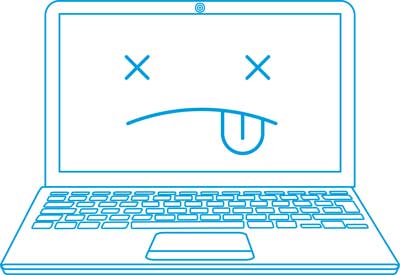
Chronic Venous Insufficiency (CVI) is a condition most commonly caused by malfunctioning (incompenent) valves in the veins. These incompent valves make it difficult for the veins to effectively send blood from the legs to the heart.
In a normal functioning vein, one-way valves open to push blood forward toward the heart, then close in order to keep the blood moving forward. When these valves do not work properly and fail to close, blood is allowed to flow back into the vein. As a result, the veins will remain filled with blood.
CVI occurs over time due to higher than normal blood pressure inside the veins of the legs. It can also be caused by Deep Vein Thrombosis (DVT) which can lead to an increase in blood pressure.
DVT is a potentially serious condition that occurs when a blood clot forms and blocks the flow of blood from the vein to the heart. Blood trying to pass the blocked vein causes an increase in blood pressure, which then overloads the valves. DVT causes leg swelling and requires immediate medical attention to prevent the blood clot from breaking off and traveling to the lungs.
Symptoms of Chronic Venous Insufficiency (CVI) may include, but are not limited to:
Risk Factors that may increase the likelihood of developing CVI:
The Venous Reflux study is used to test for valvular competence. It is performed using an ultrasound Doppler with PPG (photoplethysmography) sensor. The PPG sensor is affixed adjacent to the posterior tibial artery. While in a sitting position, the patient is asked to dorsiflex the foot a specified number of times. Dorsiflexing the foot acts as a pump for venous augmentation. Following dorsiflexion, the patient rests the foot to determine the recovery time, which is the time it takes for venous refilling to occur. Normal refill time should exceed 20 seconds.¹
The Venous Compression study involves compression of venous flow to verify augmentation, followed by proximal compression to verify valvular competence. It is performed to detect enlarged veins caused by incompetent valves. Results are interpreted by the sound produced when a Doppler probe placed over the posterior tibial vein and blood is forced out of the vein and allowed to return.
During proximal compression, a brief loud sound that rapidly concludes as blood movement is stopped by competent valves, is expected. Upon the release of distal compression competent valves will cause a rapid termination of sound (within 1 to 5 seconds).²
The Maximum Venous Outflow study uses a constricting thigh cuff to induce venous blood pooling to measure the speed with which the pooled blood flows out of the lower leg when the occluding cuff is suddenly removed. It is used to detect obstructions to venous outflow including obstructing thrombus in the calf veins, iliac veins, and vena cava. In a normal patient, the waveform will drop sharply upon release of the cuff, then rapidly return to baseline within 3 seconds.³
¹ Eberhardt, RT, Raffeto, JD. Chronic Venous Insufficiency. Circulation 2005, 111:2398-2409. Retrieved November 1, 2013 from http://circ.ahajournals.org/content/111/18/2398.
² Weiss RA. Vascular Studies of the Legs for Venous or Arterial Disease. Dermatologic Clinics, Volume 12, Number 1, January 1994. 175-190.
³ Cronenwett, JL, Johnston, KW. Rutherfords Vascular Surgery 8th Edition. Volume 1. 2014. 17:259.
© 2019 Koven Technology, Inc.